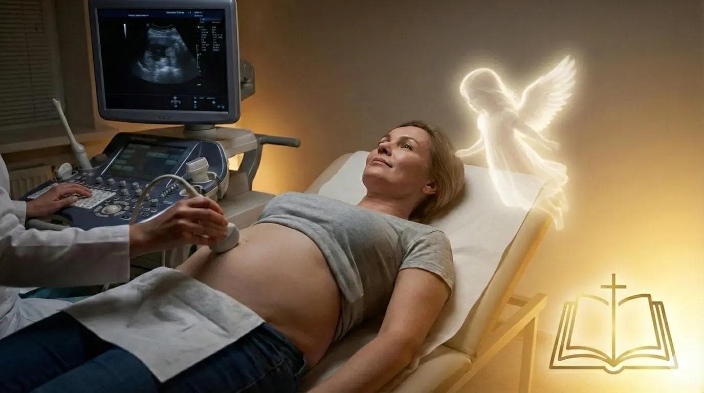
In my twelve years as a Lead Sonographer, the scent of the clinic has never changed; it is a distinct mix of sterile wipes and high-stakes anxiety. Tuesday shifts are typically reserved for baseline scans, but the chart for "Patient 402" - a pseudonym for Clara, 43 - read like a textbook case of irreversible infertility. I reviewed her history on the monitor before entering the room: elevated Follicle-Stimulating Hormone (FSH) levels consistent with menopause, an Anti-Müllerian Hormone (AMH) level below 0.1 ng/mL, and most critically, a history of a refractory thin endometrium. Her lining had measured a mere 3mm during her last cycle; for context, a viable pregnancy generally requires a "trilaminar" lining of at least 7mm to support implantation. Her Reproductive Endocrinologist (RE) had noted strictly on the requisition: "Confirm absence of follicles/cysts to proceed with cessation of treatment." We weren't looking for a baby; we were looking for the medical clearance to tell her it was time to stop trying. When I entered Room 302, the atmosphere wasn't cinematic or stormy; it was clinically heavy. Clara sat on the exam table, her demeanor flat - a common protective mechanism I see in long-term fertility patients. She wasn't weeping; she was simply resigned to the statistical reality that her body had shut down. As I prepared the 5MHz transvaginal transducer, applying the acoustic gel, I prepared my standard "compassionate but professional" script. I needed to document the state of her ovaries, measure the uterine lining, and likely confirm an atrophic uterus.
The Procedure: A Deviation from Medical Expectation
The scan began as routine. I dimmed the lights - not for drama, but to improve the contrast resolution on the high-definition monitor. In that moment, however, the clinical detachment I rely on was interrupted by a palpable shift in the room's dynamic. Patients often speak of "hope," but Clara suddenly exhibited a physiological shift; her heart rate on the pulse oximeter steadied, and her breathing deepened. She spoke of a presence in the room - a common coping mechanism in high-stress grief scenarios - gesturing toward the corner where the biohazard disposal stood. While I am a woman of science, trained to interpret grayscale pixelation into anatomical data, I have learned never to dismiss a patient’s intuition. I proceeded with the exam, inserting the probe and adjusting the gain settings to cut through the noise. I expected to see the stark, white line of a thin endometrium. Instead, the monitor displayed a complex echogenicity that shouldn't have been there. My hand froze on the controls. Deep within the fundus of the uterus, nestled in a lining that technically shouldn't have been able to support it, was a clear, circular anechoic structure. It wasn't a cyst. It was a well-defined gestational sac. I rotated the probe 90 degrees to get a transverse view, my brain scrambling to reconcile the 3mm lining measurement from her chart with the image of a decidual reaction - the thickening of the lining that occurs after implantation - blooming on the screen.
The Findings: The Intersection of Biology and Probability
I immediately switched the machine to M-Mode (Motion Mode), placing the cursor over the tiny flicker inside the yolk sac. In reproductive medicine, we live and die by the measurements, and I needed hard data before I could speak. The spectral tracing appeared instantly: a rhythmic, vertical spike pattern. I hit the 'Calc' button. Heart Rate: 168 BPM. Strong, regular, and utterly inconsistent with her hormonal profile.
When the doctor entered, he didn't gasp or cry; he went straight to the chart, flipping through the blood work pages as if looking for a clerical error. "This is a statistical outlier," he murmured, staring at the screen where the crown-rump length measured exactly 7 weeks, 2 days. He was right; medically, this was an anomaly. A spontaneous conception with an AMH of <0.1 and a history of thin lining happens in perhaps less than 1% of cases. But as I wiped the gel from Clara's abdomen and printed the thermal image, the data ceased to matter to the woman holding her breath on the table. We often tell patients that fertility is a game of numbers, but scans like this remind us that biology is not always deterministic. While we must always rely on evidence-based medicine - bolding the risks and managing expectations - there are days in the sonography lab where the physiological impossible renders itself undeniable on the screen. Clara’s "miracle" wasn't magic; it was a biological statistical outlier, reminding us that even with twelve years of experience and the best technology, life sometimes finds a way through the narrowest of margins.